- Slows spring colony population growth by 5-40%.
- Reduces honey production by 25-100%.
- Reduces pollen foraging by up to 50%.
- Fumagilin-B is essential for control of Nosema.
-
Nosema affects all aspects of beekeeping including:
- queen breeding
- package production
- crop pollination
- honey production.
Biology and effects to colonies
Nosema disease is caused by Nosema apis or ceranae, a single-celled animal belonging to a group of parasites (Microsporidia) that attack a wide-range of insects. The disease begins when spores are swallowed by adult bees. Once ingested, spores germinate and penetrate the surrounding tissues causing considerable injury to the bees’ midgut.
Bees infected with nosema experience poor digestion resulting in undernourishment and premature death. Nosema infection inhibits pollen digestion and leads to the deterioration of the feeding (hypopharyngeal) glands of nurse bees. Consequently, brood production declines.
Nosema grows and replicates in the cells of the bees’ midgut resulting in the production of millions of highly infectious spores. Ruptured gut cells release spores that spread through the gut and to other bees in the colony when voided with the feces. Spores are primarily spread through the hive by house cleaning and food sharing activities. Nosema commonly spreads between colonies through shared water sources located near the apiary.
Nosema infection can significantly reduce:
- worker life spans,
- worker nurse gland development, resulting in reduced brood rearing,
- pollen collection by workers,
- queen fertility,
- queen acceptance.
Treat with Fumagilin-B , once in fall and once in spring, for control of Nosema.
Diagnosis
There are two visible symptoms that suggest Nosema infection in a colony:
- Excessive defecation around colony entrances or on comb.
- Examination of adult bee digestive tract.
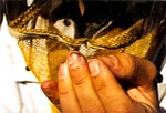
The midgut of a healthy bee is straw brown and individual constrictions are seen. Bees heavily infected with Nosema have mid guts that are white, soft, and swollen, obscuring constrictions.
|
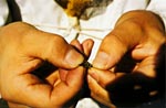
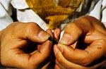
Kill a sample of 10-20 workers per colony in the freezer. Unthaw and pull tip of abdomen with figures or forceps. |
|
Pull slowly until hindgut is exposed. |
|
Healthy gut is tan to brown in color with distinct rings (left). Nosema infected hindgut whitish and lacking rings. |
Visible symptoms are not definitive indications of the disease and can be caused by agents other than Nosema. Positive diagnosis of Nosema can only be made by microscopic examination of infected bees or their fecal material for the presence of Nosema apis spores.
For microscopic confirmation of Nosema send a sample of bees from the colony suspected infected of having Nosema to Provincial or State apiarist.
Treatment